Keloid Radiotherapy: How I Do It
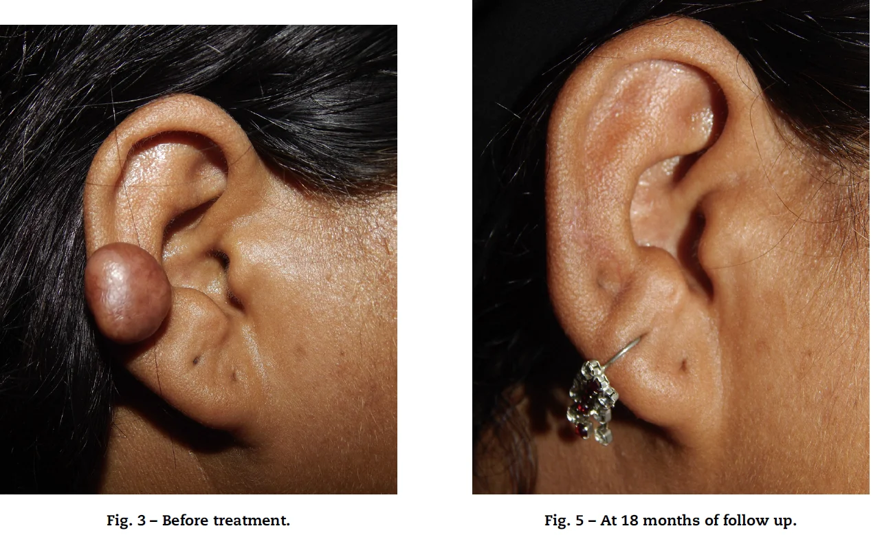
I’ve been treating around 50 - 70 keloids a year for a number of years, working with a top-class scar surgeon and using a risk-adapted radiotherapy dose schedule. I thought I’d write down some of my thoughts to stimulate discussion as this is an area which lacks good research and where there is significant variation in practice.
In this article
- Introduction
- Reading the research
- The point of this article
- 1. Establish the Diagnosis
- 2. Risk Factors for keloid formation
- Relevant Risk Factors
- 3. What are the patient’s goals?
- Common Drivers of Presentation
- A. Prominence / Bulk Is the Main Concern
- B. Desire for a Flat, Mature “White-Line” Scar
- 8. Consent – you need to include:
- 4. Surgical Principles
- Key Principles
- 5. Presenting the Post-operative Scar to the Radiation Oncologist
- Common Issues
- My practice
- 6. Timing of Post-operative Radiotherapy
- 7. Modality Selection: Matching Technique to Anatomy
- Comparing Modalities
- Depth Considerations
- 9. Dose and Fractionation
- 10. Follow-Up – An Integral Step
- Last thoughts
- References
A whole pathway approach
Introduction
I’ve been treating around 50 - 70 keloids a year for a number of years, working with a top-class scar surgeon and using a risk-adapted radiotherapy dose schedule. I thought I’d write down some of my thoughts to stimulate discussion as this is an area which lacks good research and where there is significant variation in practice.
Actually, my impression is that plenty of dermatologists and plastic surgeons don’t even consider radiotherapy in their treatment pathway and instead persist with multiple steroid injections, multiple excisions with either no adjuvant treatment or repeated (++) adjuvant steroid injections, and injections of various agents which can sometime cause a lot of tissue necrosis.
Reading the Research
Most of the literature isn’t particularly satisfying. In particular, many papers conflate hypertrophic scars with keloid scars and also treat all keloid sites equally. The best papers differentiate between keloids that are at varying risk of recurrence. I particularly admire the work of the Japanese group (Ogawa et al.) where they systematically test radiotherapy doses and changes in surgical techniques in large cohorts of patients to result in a well thought out and low recurrence rate.
The Point of This Article
So I’ve put together a bunch of recommendations, some evidence based and some that are not addressed in the literature. I’ve written it as a whole-pathway approach to keloid management, although clearly with a radiotherapy focus. Most importantly you need thoughtful and sympathetic collaboration between you and your surgeon. In this way you will reduce post-surgical recurrence rates from 80 - 100% with excision alone, or 30-40% with excision + steroid injection, to 5–10% at two years with excision + post-op RT.
1. Establish the Diagnosis
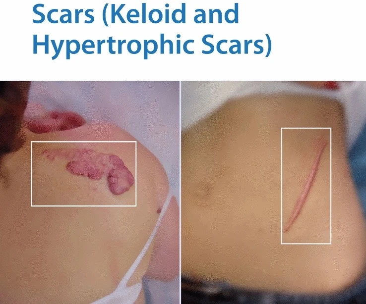
Not all scars are keloids scars! Be specific about where the scar is on the hypertrophic-keloid spectrum. Obviously there are differentials that you have to be aware of, but that’s outside the scope of this article.
2. Risk Factors for Keloid Formation
While trauma (often trivial) is the most common factor, keloid formation is often multifactorial. Many of these are modifiable, whether by GP, derm or endocrine/gynae.
Relevant Risk Factors
- Ethnicity & Family history
- Hypertension
- Smoking
- Excess weight
- Endocrine (ovarian, thyroid, adrenal)
- Follicular inflammation e.g. acne, ingrown hairs
- Location-specific anatomical tension
3. What Are the Patient’s Goals?
Patients come for various reasons, and the treatments offered should reflect their specific issues.
Common Drivers of Presentation
- Cosmetic and aesthetic
- Pain
- Itching
A. Prominence / Bulk Is the Main Concern
Some patients may be fine with flattening the scar and reducing the redness and itching. If that’s the case then the following treatments (often in combination) may be appropriate:
- Steroid tape
- Steroid injections
- Laser therapy (e.g., PDL, fractional CO₂, etc.)
B. Desire for a Flat, Mature “White-Line” Scar
If the patient wants to get rid of the scar width rather than just flattened then they need excision. I’ll talk about the requirements for effective excision below, but essentially you need to excise the whole keloid, use a tension relieving procedure, and consider adjuvant treatment.
8. Consent – You Need to Include:
- Acute and chronic skin changes
- Hair loss if appropriate
- Scar dehiscence (< 1%)
- Radiation-induced cancer
I should say here that I only treat adults. And I consider adults (generally) able to make their own decisions as long as they have all the facts. For many, the disfigurement and symptoms of a keloid outweigh a theoretical, very small risk of a skin cancer (BCC) in 20-30 years’ time. It’s a bit more tricky where the keloid overlies OAR such as breast, or thyroid and I’ve certainly come across radiation oncologists who just wouldn’t consider treating them, which is certainly not my view.
4. Surgical Principles
Surgical excision of keloids is not a generic skin procedure and apart from simple earlobe keloids I feel that it should be done by a surgeon experienced in scar management, as poor technique significantly increases recurrence risk.
Key Principles
- Excise the entire keloid
- Partial excision is associated with recurrence.
- Avoid wound tension
- Tension on the wound increases recurrence. Generally a simple ellipse excision will result in excess wound tension.
- → Use a tension-relieving closure with a wavy scar (Z/W-plasty)
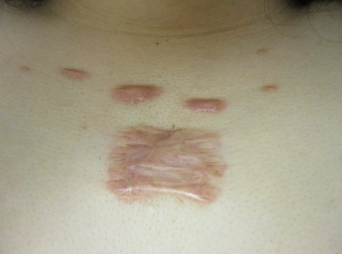
What’s interesting seeing these patients post-op is that often the scar is much bigger than you expect. For instance if you have a butterfly keloid (on the central ant chest) of 5cm diameter you may end up with a 15cm wide scar. It’s always worth asking the surgeon what they expect the post-surgical scar to look like – for instance for a butterfly keloid, knowing that the scar will overly breast tissue may significantly change the consent narrative regarding the risk of secondary breast malignancy.
5. Presenting the Post-Operative Scar to the Radiation Oncologist
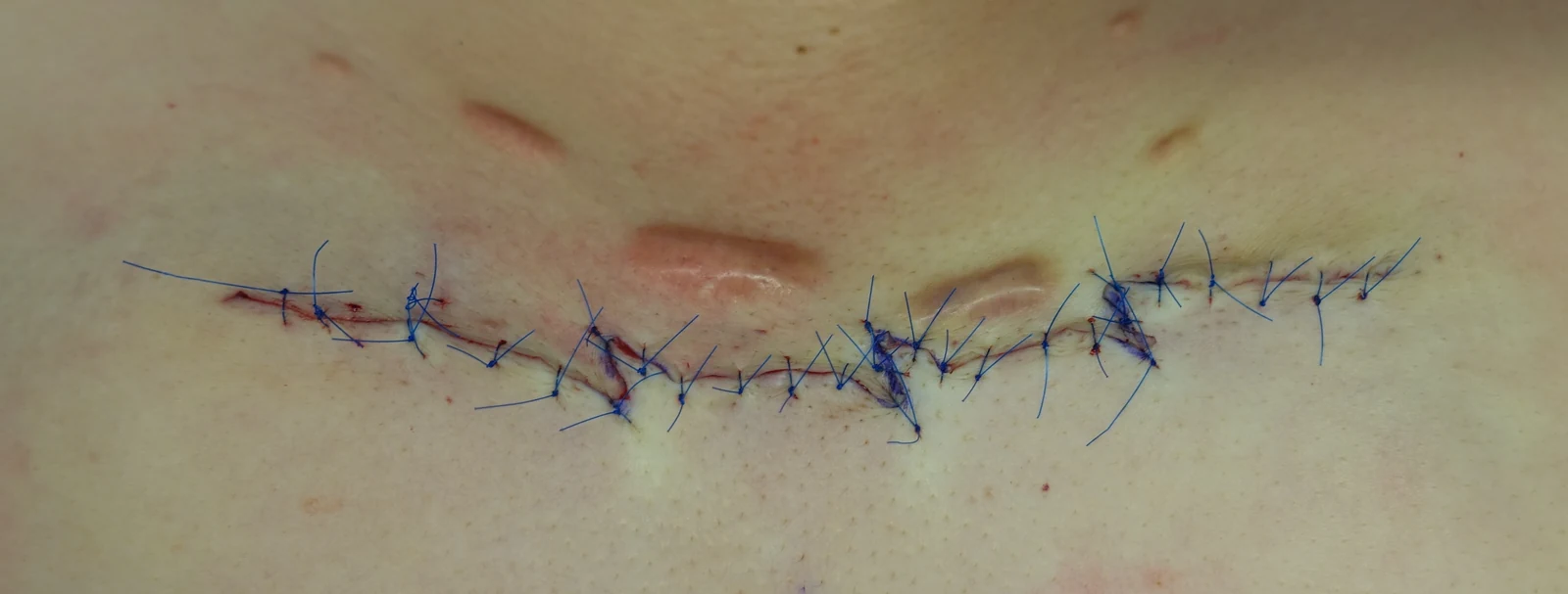
Whenever I work with a new scar surgeon (generally plastics or dermatology) I always ask them how they are going to “present” the scar. Basically I need to see all areas where the skin has been pierced during surgery, including the main scar line but also the holes where the sutures have gone through.
Common Issues
- Opaque or overly adherent dressings block visibility
- Transparent dressings may be better, but you often get bleeding that makes it difficult to see the stitch holes
- Removing dressings risks disturbing the scar and the sutures and increasing dehiscence risk
Some surgeons just leave the scar open with stitches in it, which is fine, but often the loose overlying gauze dressing adheres to the dried blood so it’s quite a job to get it free.
My Practice
My surgeon presents the scar in the following way:
- The surgeon applies an surgical adhesive tape.
- He draws the entire wound area on the tape, including the surgical track & stitch holes with an additional 1-cm margin
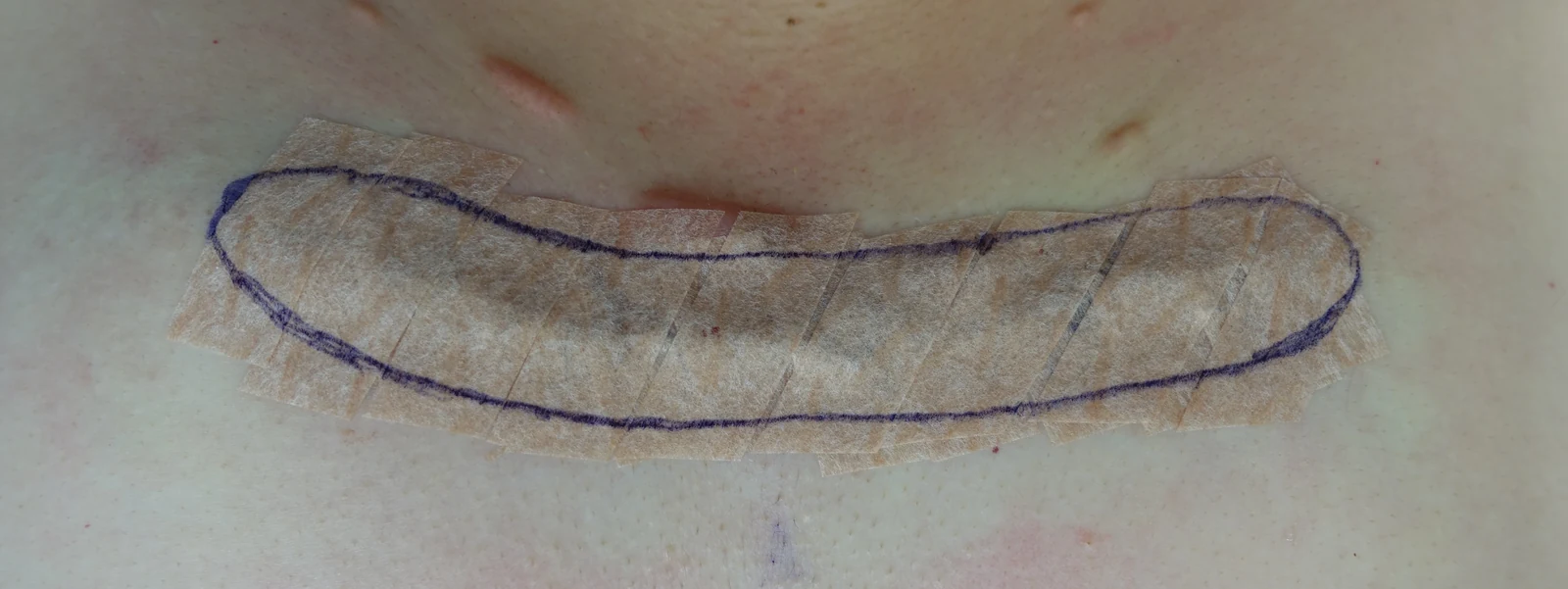
- A secondary gauze layer with micropore tape covers the area for absorption of any bleeding (to stop it bleeding on the patient’s clothes or bedsheets).
- For the radiotherapy markup session, only the outer gauze dressing is removed. The surgical tape stays in place, allowing precise field definition without disturbing the wound.
This avoids the two main pitfalls: obscured visibility and adherent dressing removal.
If I’m treating with kV photons then I simply use the field that the surgeon has marked. If I’m using electrons then I’ll add on an additional margin of 5 - 7mm all around.
6. Timing of Post-Operative Radiotherapy
Many oncologists insist on starting treatment within 24 hours, but there’s not much direct literature to strongly support this assertion, and it seems that the window probably extends to 72 hours [1-3]. Since my surgeon does his GA excisions on a Saturday I have a lot of experience with irradiating at 48-72 hours after surgery (on the Monday or Tuesday) and we have a very low recurrence rate.
7. Modality Selection: Matching Technique to Anatomy
The choice of radiotherapy modality depends mainly on what you have available in your centre. But there are some specific considerations, including lesion location (especially over OAR with regards to radiation-induced cancer risk), patient age (younger age = increased radiation induced cancer risk – does this change what you do?), treatment depth.
Comparing Modalities
- Brachytherapy
- I’ve never used brachytherapy for keloids as I’ve never had it available. Plenty of people are keen to use it due to conformality, but it’s pretty resource-intensive and just isn’t available where I am.
- kV (orthovoltage)
- I find this the easiest way to treat keloids. Quick and straightforward to plan & deliver (note – I have a conflict of interest as I’m medical director of Xstrahl who make orthovoltage machines). It’s easy to match fields if they are too long and there is not too much of an issue with curved surfaces.
- Electrons
- These give you good depth customisation but shielding is more difficult to arrange, field matching is more difficult due to the penumbra, and it results in larger fields. But until recently I only had electrons and it’s certainly possible to provide an excellent service. However, you do have to take account of:
- Curved fields - our rule of thumb is that we divide a field into two if using a single field would treat a surface of > 45 degree angle
- Field size - we have a max field size of 15 cm available so we often have to split the field in two (or even three), which results in interesting conversations about matching at depth or at the surface, under/over dosing, and feathering the junction
Depth Considerations
The aim is to cover the deep reticular dermis (i.e. the whole dermis), as this is where the keloid forming cells are. The total skin thickness is typically around 5 mm, with variation depending on body site (see the Oltulu paper for details [4]), so I aim to cover a depth of at least 5mm and often up to 10mm, depending on body site.
There are some areas where the consideration is really about separation of the superficial and deep aspects of the volume, for instance when you treat an earlobe, where you may have to cover e.g. 1.5 - 2cm including dressing/swelling.
9. Dose and Fractionation
My practice follows the Japanese risk-adapted scheme [1]. It’s derived from a baseline regime of 15Gy in 3 fractions over 3 days. They started off by using that adjuvant dose for all keloids in 249 patients up to 2002. They found recurrence of:
- Overall = 30%
- Highest = 40% (chest wall, scapula, suprapubic, auricle)
- Lowest = 6% (earlobe)
From 2003-2012 they changed the number of 5Gy fractions to 20Gy in 4# for high-recurrence sites, 10Gy in 2# to earlobes and kept 15Gy in 3# for ear auricle. That gave them an improved overall recurrence rate of 12% in 644 patients (compared with 30% before) and improvement in high-risk sites to 13%, maintenance of low recurrence at earlobes, but still quite a high auricle recurrence rate.
- Overall = 12% (was 30%)
- High rec sites = 13% (was 40%)
- Auricle of ear = 21% (was 40%)
- Earlobe = 7%
Meanwhile, from 2008-2012 they improved their surgical techniques:
- Earlobes - wedge excision, simple superficial sutures: Rec Rate 6.9% → 4.6%
- Ear auricle – core excision (not wedge): RR 21.4% → 9%
- High rec sites (Chest Wall, Scapula, Suprapubic)
- Excision, subcutaneous/fascial tensile reduction sutures and z-plasties
- Used for most pts throughout whole 2003-2012 period, so no change in RR
Then they changed the RT protocol in order to reduce the number of fractions as follows:
- Earlobe = 8Gy/1#
- Auricle = 15Gy/2#
- High recurrence sites = 18Gy in 3 fractions
And recurrence rates in 494 patients were:
- Overall = 9.3%
- High-rec = 10.4%
- Auricle = 4.5%
- Earlobe = 6.7%
So that’s the dose-fractionation scheme I use. Totally understand that different people have their own favoured recipes, but there are such a large number of patients in this study and it makes sense to me to treat different sites in different ways. I quote a 5 - 10% recurrence rate at 2 years, as most keloids will have recurred by then.
10. Follow-Up – An Integral Step
Follow-up is often overlooked. Many patients are discharged at 3 – 6 months after surgery, which can miss recurrence. And the point is that early scar hypertrophy can be salvaged. So we follow up 3 monthly for at least a year. And if there is early thickening then our practice is to start with a steroid plaster and then to move onto steroid injections.
By the way, this raises a more basic issue - what do we actually mean by recurrence?
- Is a clinical diagnosis enough or do you need pathology?
- Does the scar have to get to the pre-surgical size or just grow a bit? How about widening and a bit of thickening of the scar?
- How about if it grew post-RT and then was salvaged – was this failure that was salvaged or was it, by definition, never failure in the first place?
Last Thoughts
I’ve given my own personal thoughts on adjuvant keloid radiotherapy. I’ve purposely steered away from primary radiotherapy, which I don’t really use much, although I think it’s an interesting and under-researched area. I’ve also not covered post-RT recurrence, about which there is a similar lack of literature.
I’d be interested in your comments - lets discuss!!
References
- Ogawa et al. Surgical excision and postoperative radiotherapy for keloids. Scars, Burns & Healing Volume 5: 1–11. DOI: 10.1177/2059513119891113
- Kovalic et al. Radiation therapy following keloidectomy: A 20-year experience. IJROBP 17:77-80
- Lo et al. Single-dose electron beam irradiation in treatment and prevention of keloids and hypertrophic scars. Radiotherapy and Oncology 19 (1990) 267-272.
- Oltulu et al. Measurement of Epidermis, Dermis, and Total Skin Thicknesses from Six Different Body Regions with a new Ethical Histometric Technique. Turk J Plast Surg 2018;26:56-61.
Next steps
Thinking About Treatment for Keloids?
Dr Shaffer sees patients for assessment and, where suitable, low-dose radiotherapy.